Last Updated on March 29, 2022 by Allison Price
Equine recurrent eye disease (ERU), a serious ophthalmic condition, is reported to be 2%-25% in the world . ERU classic is characterised by periods of active intraocular inflammation that are followed by variable quiescent times. Some horses may experience subclinical ocular inflammation (ERU) that persists indefinitely without any obvious discomfort. Chronic inflammation can lead to secondary ocular conditions such as cataracts and lens luxation. ERU is therefore the leading cause of blindness among horses worldwide.
Equine Recurrent Uveitis: Etiology and Pathogenesis
Equine recurrent eyeitis (ERU) is an autoimmune condition that occurs following an episode of acute uveitis. ERU can occur in horses who have had a single episode of acute uveitis. However, not all horses will be affected. ERU can be initiated by many causes including viral, protozoan and parasitic. However, the pathophysiology is multifactorial and complex. Leptospira spp were the most studied for their role in initiating ERU. Although the exact mechanisms by which Leptospira people initiate ERU are not known, it is probable that loss of ocular immuno tolerance, cross-reaction between Leptospira organisms, self-antigen, as well as intra- and intermolecular Epitope spreading, all play a crucial role.
ERU is not predisposed to any gender or age. ERU horses with Appaloosas and Warmbloods are more likely to be than horses with . This suggests that there is a heritable component. The major histocompatibility and/or leptospirosis genes (MHC) have been the most extensively studied and are likely to play a role. ERU prevalence varies by geographic location. higher rates are reported in temperate and tropical climates than in dry, arid climates . This observation may partly be due to variations in the persistence of pathogenic Leptospira species within the environment. ERU development is dependent on the causes of primary uveitis. These factors include the genetic makeup of horses and the environment.
Clinical Findings and Lesions for Equine Recurrent Uveitis
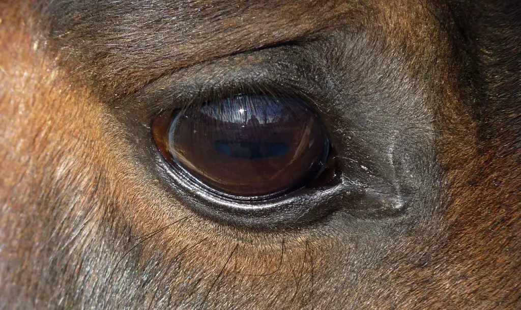
Equine recurrent eye problems can be characterized by both acute and chronic secondary complications. Leukocytes can invade the uvea by releasing proinflammatory cytokines like prostaglandins or leukotrienes due to changes in ocular immunity. These mediators increase vascular permeability in the uvea and cause breakdown of the blood-ocular shield, iris muscle spasm, decreased production of aqueous humor, and ciliary muscle spasm. These changes cause the classic symptoms of acute uveitis, including epiphora and blepharospasm as well as corneal edema and episcleral congestion. The anterior segment signs can often make it difficult to see the posterior segment. Vitreous haze can be caused by active posterior segment inflammation due to cellular infiltrates, fibrinous bands, focal or diffuse retinal detachedment or chorioretinitis.Active uveitis, horse
COURTESY OF UC DAVIS VETERINARY OPHTHALMOLOGY SERVICE.Equine recurrent uveitis
COURTESY OF UC DAVIS VETERINARY OPHTHALMOLOGY SERVICE.
ERU Chronic sequelae include:
- Corneal scarring
- iridial fibrosis
- corpora nigra atrophy
- posterior synechia
- Cataract
- lens dislocation
- glaucoma
- Phthisis bulbi
- Chorioretinal scarring
ERU can affect the eyes in asymmetrical ways.
Primary ocular disease in horses, particularly corneal disease, can cause many of the same symptoms as ERU. These include epiphora, blepharospasm and miosis. It is crucial to distinguish primary corneal disease and ERU due to the differences in treatment. An thorough ophthalmic exam and fluorescein stain is essential. Prepurchase and soundness examinations require a thorough ophthalmic exam, which includes fundoscopy. Horses suffering from chronic ERU may not show any anterior segment signs, but they could have severe retinal degeneration. This can lead to vision compromise.
Diagnosis for Equine Recurrent Uveitis
- Examen of both eyes in detail
- Tonometry
- Fluorescein stain
Equine recurrent eyeitis can be diagnosed by looking at the clinical signs and the history of persistent or recurrent episodes. To rule out other primary ocular diseases and observe signs consistent with ERU, a thorough ophthalmic exam of the anterior and posterior segments is essential. To rule out glaucoma, and to document hypotony associated with ERU, it is important to perform Tonometry in all cases. Fluorescein stain can be used to evaluate corneal epithelial integrity, and exclude reflex uveitis due to ulcerative keratitis.
A physical exam is required in cases of severe uveitis to rule out other conditions. As part of the minimum database, a CBC and a serum biochemistry panel may be necessary. Specific tests can help determine the cause of primary uveitis. While serologic testing for Leptospiraspp may confirm that you have been exposed to this risk factor, it is not able to determine the treatment. Although anterior chamber paracentesis or vitreous cavity parocentesis can help to identify the causative organism, it is not recommended as this may result in severe intraocular damage.
Equine Recurrent Uveitis: Treatment, Prevention and Control
- To reduce inflammation, topical steroids and nonsteroidal anti-inflammatories systemic are combined.
- As an adjunctive treatment, topical atropine can be used to reduce intraocular pain or cause mydriasis.
- To reduce the number of uveitic episodes in visually impaired patients, a suprachoroidal implant can be made or vitrectomy may be performed.
- Blind, painful eyes should be treated with enucleation.
Therapy for equine persistent uveitis has three main goals: to reduce inflammation, alleviate discomfort, and prevent vision impairment. The specific cause of the underlying condition should be identified and treated as part the initial treatment plan. To minimize intraocular inflammation and regardless of the cause, aggressive treatment with topical and systemic anti-inflammatory medication should be initiated immediately. The initial treatment of acute uveitis in horses is crucial. Flunixin meglumine should be administered systemically (especially IV). Initial IV doses are typically 1.1 mg/kg. This is followed by a 5-to-7-day course of 0.5-1 mg/kg PO twice daily. The dosage can be decreased to 0.25 to 0.5 mg/kg every other day or once daily as the inflammation subsides. This is for a 1-to 3-month treatment period.
Flunixin meglumine can cause renal toxicity. Therefore, it is important to monitor serum creatinine every other day if flunixin is administered for more than one month. Flunixin meglumine-treated horses should be monitored for signs of GI ulceration. Concurrent prophylactic administrations of omeprazole (2 mg/kg/day PO) may also be recommended. Flunixin meglumine can be tolerated if it is not. However, aspirin (10-25mg/kg PO, once or twice daily) and phenylbutazone (2-4 mg/kg PO) may also be used. Horses with chronic, low-grade uveitis or frequent recurrences were treated medically by taking daily or every other day aspirin or oral phenylbutazone. Most horses can tolerate this medication well. However, adverse GI, hematologic or renal effects may occur and the regimen is not always effective in eliminating recurrence.
Prednisolone (100-300mg/day) or dexamethasone (5-10mg/day) can also be used to treat acute episodes of uveitis. However, their long-term use has been linked with laminitis. Systemic antibiotics should not be used unless there is a bacterial infection.
Topical steroids, such as dexamethasone (1% suspension or ointment), and prednisolone Acetyl (1% suspension), can be very effective in decreasing inflammation. Topical steroid preparations and suspensions are better than sodium phosphate formulations because they penetrate the cornea and reach adequate uveal concentrations. Topical hydrocortisone is to be avoided as it does not penetrate the cornea well enough and is not potent enough to treat anterior uveitis.
Fluorescein staining is required before starting topical steroids. These medications are contraindicated for corneal ulceration or infection. Topical nonsteroidal medications such as flurbiprofen (0.3% solution) or diclofenac (0.1% Solution) are less powerful than topical steroids, but provide a greater safety margin for patients with concurrent corneal disease. The severity of inflammation determines the frequency of administration. Initially, it may be administered 4-6 times per day. The frequency of nonsteroidal and topical steroids can gradually be decreased as clinical signs improve. After active inflammation has subsided, treatment should be continued for 1 month.
Topical atropine (1% solution, ointment), causes mydriasis and cycloplegia. It stabilizes the blood-aqueous boundary and decreases the possibility of posterior synechia development. The pupil should be dilated by applying atropine topically 2 to 3 times per day. Once the pupil has dilated, the frequency can be adjusted to maintain mydriasis. Horses treated with topical atropine need to be closely monitored for signs and symptoms of ileus, as atropine reduces GI motility.
Subconjunctival injections with triamcinolone Acetamide (1-2 mg), provide intraocular anti-inflammatory levels for 7-10 days. They are less likely than other steroids (10-40 mg) to cause abscess formation or granuloma formation. Subconjunctival steroids must be used with caution. They cannot be removed once they are injected.
There are two surgical options that can be used for long-term management. A suprachoroidal-cyclosporine cyclosporine insert is a sustained release medication device that delivers therapeutic levels of cyclosporine, an immunosuppressive, T-cell inhibitor. It lasts approximately 3 years. The procedure involves the placement of a cyclosporine-A disk measuring 5 mm in size under a scleral flap that is placed 8mm posteriorly to the dorsolateral side of the limbus. Implanted horses have significantly fewer episodes of uveitis than before surgery. This device also provides effective long-term control of ERU.Horse Suprachoroidal Cyclosporine Implant, horse
COURTESY OF UC DAVIS VETERINARY OPHTHALMOLOGY SERVICE.
Core vitrectomy involves removing virtually all vitreous via an incision just posterior to the dorsolateral side of the limbus. The vitreous can then be replaced with either balanced salt solution, or saline. This procedure has the potential to reduce chronic inflammation caused by organisms such as Leptospiraspp and/or inflammatory cell in vitreous. These factors can be eliminated to reduce the severity and frequency of uveitis episodes. Eyes that are either blind or pain from ERU should be considered for enucleation.
ERU can be managed with good husbandry practices. This will ensure that the animal is healthy, avoids ocular trauma and minimizes environmental triggers. Routine deworming, vaccinations, proper nutrition, and dental care are all recommended. These measures may be beneficial for individual horses but they have not been assessed to determine how they affect the clinical course and outcome of ERU.
The Key Points
- Recurrent uveitis in horses can lead to blindness or ocular pain.
- For vision and comfort, prompt diagnosis is combined with medical and surgical management.