Last Updated on March 15, 2022 by Allison Price
Horses are prone to liver disease. It can be difficult to determine the cause of liver disease, despite this being a common problem in horses. The main organ responsible for the metabolism, accumulation and distribution of nutrients in the body is the liver. Most nutrients that are absorbed through the gastrointestinal tract travel directly to the liver via the portal circulation. There they are metabolised and repackaged, then stored or exported to other tissues. The liver plays an important role in detoxification, both of endogenous and exogenous substances. All toxic substances that enter the gastrointestinal tract through the stomach are passed to the liver, which acts as the first line of defense. Many substances absorbed into horses’ diets can cause liver damage. Inflammation of the intestines and gastrointestinal disease can also cause liver injury.
Below is a table that lists some possible causes of liver disease in horses:
| Toxins | Plants (e.g. Ragwort, clover)Mycotoxins (e.g. aflatoxin, zearalenone, fumonisin) Chemicals Drugs |
| Infections | Viral hepatitis (Equine herpes virus, Equine parvovirus hepatitis, Equine hepacivirus) Parasitic (Liver fluke, large strongyles, ascarids) Cholangiohepatitis |
| Inflammatory | Chronic active hepatitis Secondary to gastrointestinal diseases |
| Other | Neoplasia Cholelithiasis Hyperlipaemia |
Clinical Signs & Diagnosis
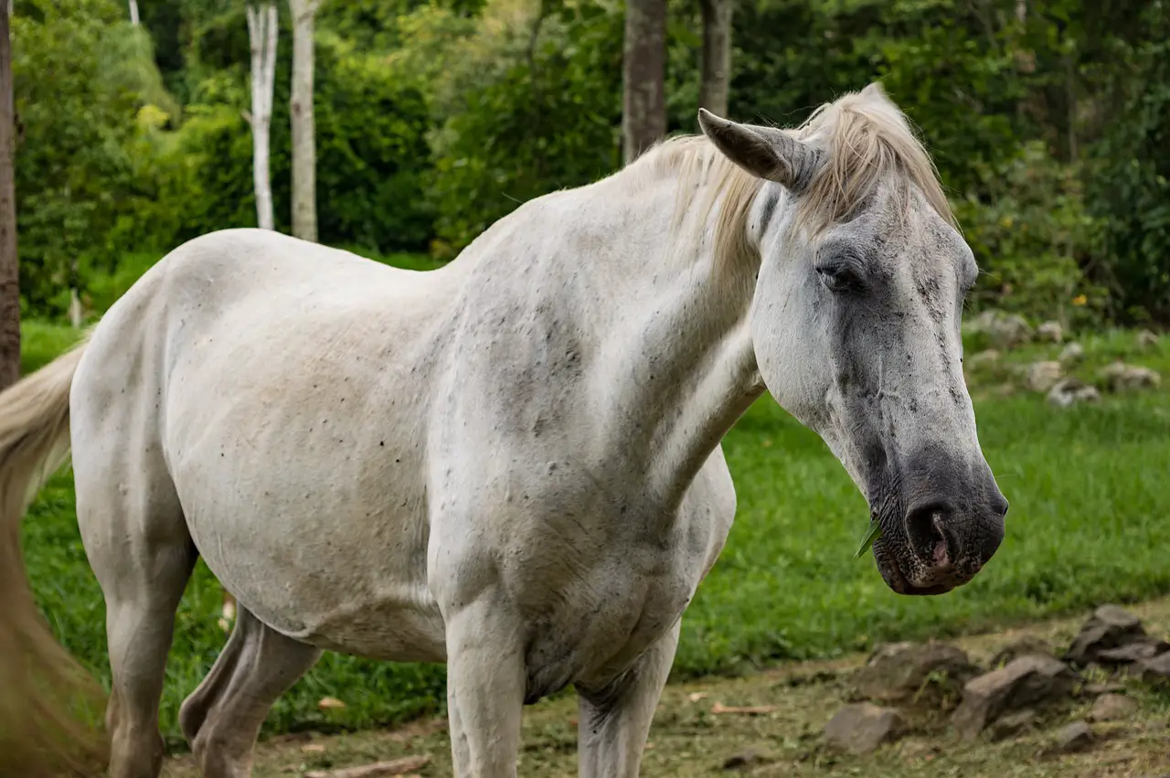
In the early stages of liver disease, signs are often vague or non-specific. Low levels of hepatic injuries in horses are not always obvious. Weight loss, anorexia and colic can all be signs of hepatic injury. The main image depicts a horse suffering from end-stage liver disease and severe oedema in his abdomen.
Biochemistry can be used to quickly diagnose liver disease or injury. This will usually reveal an increase in the concentrations of hepatic enzymes such as gamma glutamyltransferase, glutamate dehydrogenase and aspartate aminotransferase. Analyzing bile acid or bilirubin levels can provide additional information on hepatic function. Other laboratory findings that may be found in horses suffering from advanced hepatic diseases include increased blood clotting time, hyperglobulinemia, and an increase in ammonia.
After determining that the horse is suffering from hepatic disease, or an injury to his liver, further diagnostic tests may prove useful in identifying the root cause. It is important to determine if the affected horse is an individual or a group. It is simple to test blood from several horses in the same environment. It is common to find other horses subclinically affected. A group of horses with a similar condition could indicate that they have been exposed to an environmental toxin or other infectious agent.

A horse suffering from liver disease may benefit from hepatic ultrasound. This allows for the assessment of liver size and parenchyma. It can also identify specific problems like choleliths, dilated or blocked bile ducts, and masses. These changes can be subtle and non-specific, and are common in most horses. No specific abnormalities have been observed. To provide additional diagnostic and prognostic information, hepatic biopsy may be performed. Ultrasound guidance should be used for this purpose. Histology can be used to diagnose specific conditions (e.g. ragwort toxicity or neoplasia), but it does not always identify the aetiologic agent. However, it provides valuable information about the disease process and severity. This information can help to develop a treatment plan and give prognostication.
It can be difficult to identify potentially toxic substances. Visual inspection of the pasture and hay for signs of toxic plants like ragwort is a good first step. To determine if there are other unusual toxins, such as arsenic and other heavy metals, it is important to take a detailed history. If necessary, specific testing can be done.
Mycotoxins can cause liver disease in horses. This is a serious and often overlooked problem. Mycotoxins are toxic substances produced by moulds that grow on crops during harvest, storage or growth. The production of toxins can be affected by environmental conditions. These substances are more likely to be produced in times of stress, such as extreme temperatures or dry weather. These toxins can survive for long periods on pasture/forage once they are produced. They can also be metabolized by acid in the stomach. Rossdales Laboratories has teamed with Alltech who are the world’s leaders in animal nutrition management and mycotoxin management to offer mycotoxin testing. This involves screening feed samples (usually forage) to determine if there are any mycotoxins. This allows for a risk analysis to determine the likelihood of developing a mycotoxin-related disease.
A number of new viruses have been linked to liver disease in horses in recent years. Equine Parvovirus-Hepatitis (EqPV–H) has been recognized as the cause of Theiler’s disease (equine sepsis hepatitis), which is often but not always associated with recent administrations of an equine biologic drug ( Divers and al, 2018 ). The most common signs of clinical disease are lethargy and anorexia. Although the mortality rate for clinical cases is high due to high rates of infection, horses infected with subclinical hepatitis can present with elevated levels of hepatic enzymes and may have subclinical symptoms. Equine Hepatitis V (EqHV) was also recently discovered. It is closely related with the human Hepatitis B virus. Although research into the role this virus plays in horse liver disease is ongoing and may cause mild elevations of hepatic enzymes, it could also be linked to subclinical disease in horses. EqPVHV and EqPVHV can both be detected in horses that are clinically healthy. These horses may be carriers. If a biopsy is performed, PCR analysis can detect both EqPV-H and EqHV in serum samples or liver tissue.
Parasitic infected can cause focal or multifocal hepatic diseases, but it is rare to cause liver disease or severe hepatic dysfunction. Large strongyles and larvae of Parascaris can enter the liver and cause focal fibrosis and hepatitis. Echinococcus Granulosus may cause hydatid cysts within the liver. These can be seen on ultrasound, but they are often an accidental finding. Horses can get liver fluke infection, especially if they are co-grazing with other ruminants. However, it is more common to cause subclinical disease. Although fecal testing for fluke eggs can be done, it is not sensitive due to intermittent egg shed and the fact many infections don’t become patent. A serological test can also be used to aid in diagnosis.
Primary bacterial hepatitis/cholangiohepatitis is rare in adult horses. Cholangiohepatitis may occur secondary to bile stagnation (e.g. Cholangiohepatitis can also occur secondary to bile stasis (e.g., cholelithiasis or intestinal obstruction). The diagnosis is made based on histopathological findings from liver biopsy and the isolation of bacteria from fresh tissue samples. Most commonly, cholangiohepatitis is caused by bacteria isolated from the small intestine.
Cholelithiasis is a rare condition that occurs in older horses, and may be associated with bacterial cholangiohepatitis. This could predispose to cholelith development. Multiple choleliths are common in cholelithiasis. Cholelithiasis may be present at any level of your biliary system. Choleliths can be silent in the clinical setting or cause colic and hepatic disease. Ultrasonographic findings may include increased hepatic echogenicity and cholelith visualisations. Histopathological findings and hepatomegaly can also be helpful.
Primary Hepatic Neoplasia is extremely rare in horses. Metastatic spread (e.g. Although lymphoma and malignant melanoma are more common, it is still rare. An antemortem diagnosis may include ultrasound, biopsy and histological examination.
Summary
It can be difficult to diagnose liver disease in horses. The first step is to take a thorough history and perform a physical exam. Each case is different and may require further investigation, such as blood testing, ultrasound, or biopsy. Horses that are affected in a group should have mycotoxin testing. You can also use viral testing or liver fluke serology to test for other diseases.