Last Updated on March 23, 2022 by Allison Price
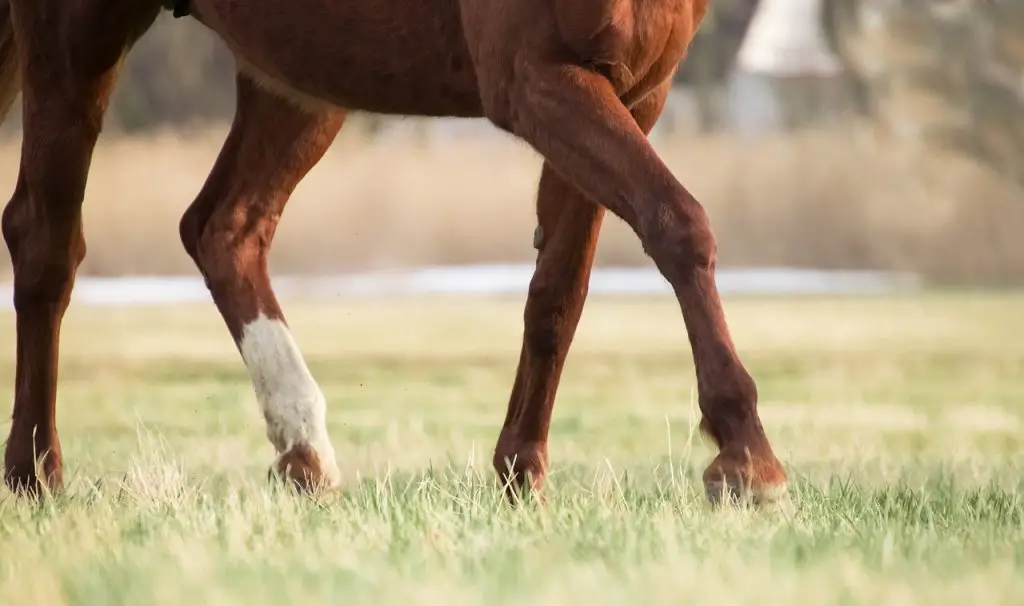
Distal Tarsitis is the most common cause for clinical lameness in horses due to the tarsus (or “hock”) of the horse. Distal tarsitis refers to osteoarthritis, periostitis, and tarsometatarsal joint inflammations.
What is the distance between the distal and tarsal joints?
The structure of the tarsus (or “hock”) is very similar to that of the human heel. The tarsus is made up of four joints: the tibiotarsal, proximal, distal, and tarsometatarsal joints. The tibiotarsal joints perform approximately 98% of the motion in the tarsus. Just below the tibiotarsal joints, the proximal-intertarsal joint performs about 2% of the motion. The distal intertarsal joint and the tarsometatarsal joint, which are at the bottom of the motion spectrum, move very little. These joints would not be present if the horse didn’t have them (i.e. If there were no solid bones across the distal torsus, we wouldn’t be able detect any abnormalities or changes in gait.
How can distal tarsitis occur?
The distal tarsal joint is not necessary for normal horse locomotion, but they can be unstable. Joint inflammation, also known as arthritis, can develop from chronic joint instability (i.e. arthritis). Distal tarsitis can be caused by repeated compression and rotations of the tarsal bone and excessive tension at the attachment of major dorsal ligaments. The following factors can influence the development of distal-tarsitis: age, weight, breed and job description of the horse, as well as the frequency, intensity, and type of work done.
These are some conformational abnormalities that could increase horse’s chance of developing distal Tarsitis.
- Straight pelvic limbs
- You can also make hocks.
- Cow hocks
What are the clinical signs of a syphilis?
Horses suffering from distal tarsitis often experience gradual onsets of pelvic limb weakness. Lameness can be most evident during trot. It may also be characterised by hypermetric “stabby” pelvic limb flight patterns. Horses will often pull their pelvic limbs under their bodies and “stab” them as soon as the foot touches the ground. Horses will prefer the affected limb when exercising, even though lameness can be bilateral. A horse may show a “hip hike” (pelvic excursion), when they are trotting with one limb affected. After a rest period, lameness can worsen. Affected horses often exhibit stiffness when they first start to exercise, but are sometimes able to “work it out”. If the condition is chronic, a firmening of the hock can be seen. This is due to excessive bone growth at the distal tarsal joint. Horses suffering from moderate to severe tarsitis are likely to show a positive Churchill’s Hock Test. This procedure is done during the passive lameness assessment. The pelvic limb abduction is a sign of a positive response. Spavin testing, which is performed during active lameness evaluation, is a reliable and widely-used method to detect distal tarsitis. After 60-90 seconds of pelvic leg flexion before trotting, a positive response will result in hypermetria and increased lameness.
Horses can develop secondary symptoms if they are prone to experiencing side effects from having one or both of their pelvic limbs favor for a prolonged period. These are common compensatory issues:
- Thoracolumbar ebaxial soreness (back) due to an asymmetrical pelvic limb gait
- Chronic overloading of the thoracic spine limbs can cause proximolateral thoracic leg suspensory desmitis
- Greater Trochanteric bursitis (“whorlbone”) is a result the abnormal pelvic limb gait
- In an effort to alleviate hock pain, there is increased wear on the outside side of the shoe or pelvic foot.
These abnormalities are often secondary or unrelated to distal tarsitis. Often, only successful treatment of the problem can resolve it. Horses with any of the clinical signs mentioned above should be examined for distal tarsitis.
How can distal tarsitis be diagnosed?
Distal tarsitis can be diagnosed clinical. In other words, distal tarsal pain is diagnostic. Clinical examination, lameness characteristics and the response to Churchill’s Hock Test, hock flexion and intra-articular injections are all indicators of pain. To assess the severity and presence of distal tarsitis, radiographs are often used. However, radiographs do not provide structural information and cannot show joint inflammation (arthritis). Radiographic changes and distal tarsitis are not always correlated because the tarsus has low motion. Because it provides functional information, nuclear scintigraphy (bone scan) might be more reliable in determining the presence of distal-tarsal inflammation. Measures bloodflow. This diagnostic method has proven to be extremely useful in the diagnosis of distal tarsitis.
How can distal tarsitis be treated?
There are two basic types of treatment for distal tarsitis. The first is to reduce and/or eliminate inflammation in the distal tarsal joint. This can be achieved through intra-articular and systemic anti-inflammatory therapy. Comfort is improved by reducing inflammation (arthritis). Steroids are often used in intra-articular therapy. They can be very effective at reducing pain and inflammation. This treatment also aims to preserve normal synovial integrity in the distal tarsal joint. Adequan (r), Legend (r), Cosequin (1r), and others are systemic medications. These medications are intended to improve the horse’s synovial function as well as general joint comfort. The Atlanta Equine Clinic uses intra-articular Hyaluronan Therapy (in combination with steroids), to increase the local effect.
Fusion of the distal intertarsal or tarsometatarsal joint is another form of distal-tarsal therapy. This can be done surgically or with a chemical agent that is infused into distal tarsal joint. Because these joints are almost motionless, fusion causes minimal movement to the horse’s gait. However, the joint can be removed to eliminate instability, inflammation, and pain. This is usually reserved for horses who have failed to respond to anti-inflammatory treatments and have experienced osseous changes related to the distal tarsal joint. Ask one of our staff for more information about distal-tarsal fusion. We are happy to talk more about this topic.