Last Updated on March 31, 2022 by Allison Price
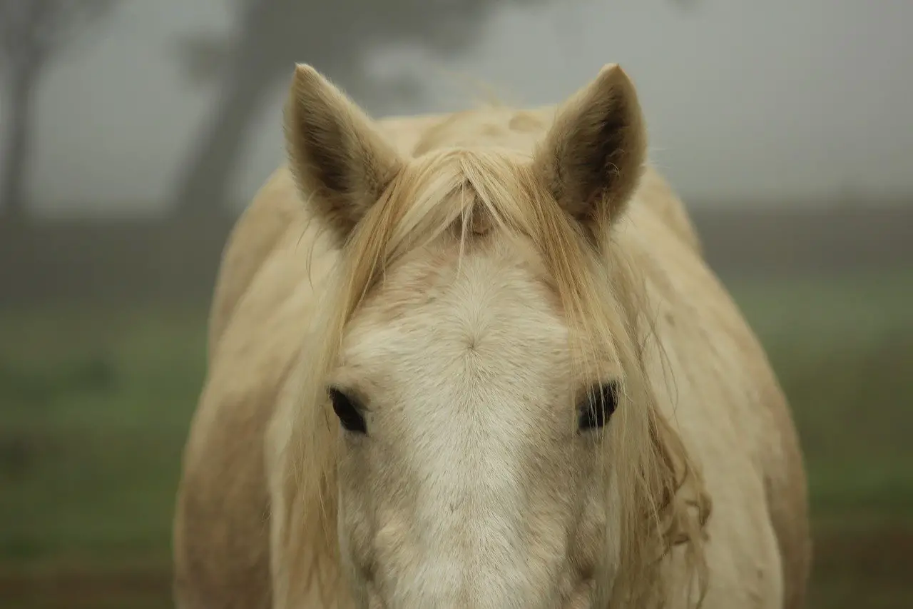
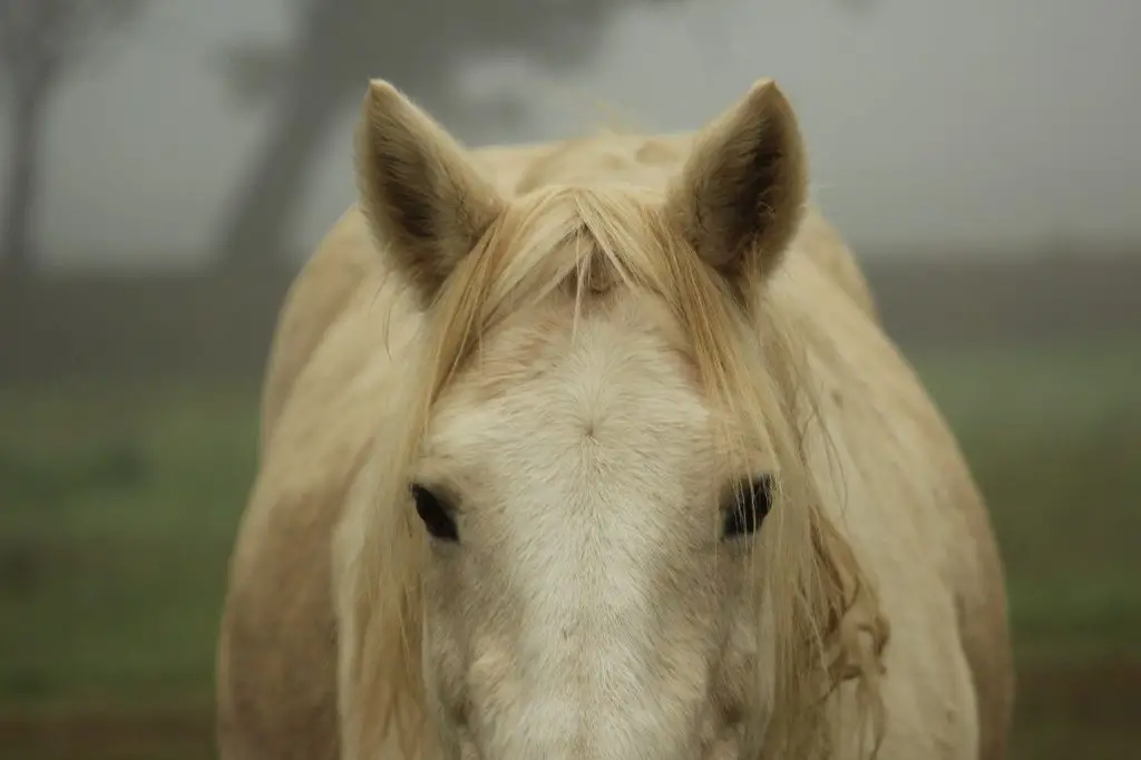
Melanoma, a common skin condition that causes nodules in older grey horses, is very common. It usually occurs between 7-8 years old. Over 80% of grey horses will experience at least one form of melanoma in their lifetime.
Melanoma can occur in horses at any age, some even at birth.
Many people believe melanomas in grey horses are benign skin tumors. Although most melanomas are benign, they can develop into malignant. Their location can have serious consequences for horses welfare.
Malignant melanoma will develop in more than 80% of cases. Some melanoma lesions will become malignant quickly, while others may remain benign for a longer time before becoming malignant.
Horses with melanomas that are not grey tend to be more serious. They usually occur as a single, isolated lesion. They are more common in grey horses and can be found in large numbers.
It doesn’t matter how large the tumor is. Sometimes small melanomas are malignant, while larger ones can be benign.
Melanoma in horses can progress to the point of being fatal.
What horses are affected?
It is possible that the breed susceptibility to melanomas is not entirely true. The most important predisposing factor is the colour grey.
Grey horses aged between 10-12 years will likely have at least one melanoma lesion. Many horses with multiple lesions are affected.
The susceptibility to melanoma in horses that are flea-bitten grey with brown or chestnut flecks appears to be lower. These colours are still more susceptible to melanoma than chestnuts and horses of other colors.
Non-grey horses can also be affected by melanoma, while cremello/albino horses may be more severely affected. Melanoma in non-grey horses is more serious than it is in grey horses.
The Percheron, Arabian, Lipizzaner and Andalusian breeds seem to be more vulnerable, while the naturally grey Erisakay pony seems to be less affected.
What is a Melanoma?
Melanoma tumors are easy to identify. Melanomas can be described as small, isolated nodules that have a dark colour. Melanomas do not regress spontaneously.
Melanomas often begin as small, solid, spherical masses under the skin. They can form chains or vary in size. While one tumour might grow rapidly, others may not. They can become more dangerous and grow larger, causing them to merge into larger areas.
Lesions that grow faster and are damaged or injured more easily may develop ulcers and expose black soft tissue. Melanomas can bleed and produce a black tarry jelly-like substance.
Sometimes melanomas lose colour and become grey/blue, then beige, and finally red/pink.
Other cutaneous nodular diseases, such as carcinoma and sarcoid, can mimic melanoma or lead to misinterpretation. Melanoma’s clinical appearance is so distinct that tissue sampling is not usually necessary for histological confirmation. However, samples from the suspected melanoma will often be confirmed by either a needle or an excision of the tumour.
Where do Melanomas occur?
Melanomas occur in the skin, ear and eye. The cells responsible for them (melanocytes) are normally responsible to the colour of the skin and therefore most lesions are found in the skin.
If a melanoma is found internally it is due to spread from a malignant melanoma of the skin
The most common site for melanoma is the skin of the perineum (around the anus and the base of the tail). More than 50% of melanoma tumours are situated in and around the perineum.
Melanoma can also occur in the following areas:
- Eyelids, iris and retina
- Mouth, in particular the lips
- Parotid salivary glands and lymph nodes
- Penile and vulval skin
- Internal organs, including the intestine, heart and lungs.
Melanoma may have little effect on an individual organ until it reaches a size that interferes with the function of that organ, however in some sites such as the eye and the spinal cord, even small tumours can have a devastating effect.
What causes Melanoma
Melanoma in humans is linked to sunburn and exposure to sunlight. Grey horses are more likely to develop melanomas in areas that have not been exposed to too much sunlight. While we do know that horses are susceptible to melanomas due to their genetic makeup, very little is known about the causes.
Melanomas usually start with one-cell changes that cause an expansion of abnormal cells. An abnormal accumulation of melanin pigment within otherwise healthy cells seems to be the trigger for cancerous changes. The abnormal and prolonged presence of melanin pigment in cells appears to trigger changes to the cells’ genetic structure.
These “tumours”, which appear in the early stages, are likely to be very benign.
Non-grey horses, dogs and humans may develop melanomas after exposure to ultraviolet light or other cancer-inducing factors.
What does Melanoma do to the horse?
The main effects of melanoma are their location and the changes they make to the tissue structure. The severity of the effects will depend on their size and location. Even if the tumour is not malignant, it can have serious clinical consequences. A heart attack or melanoma can lead to severe irregular heart rhythm problems that could cause sudden death.
Massive melanoma can occur in the perineal area and may affect defaecation or urination. Melanomas in mares can cause reproductive problems with mating and parturition.
Melanomas can cause irreparable damage to the local anatomy of various organs. This problem is most commonly found in the region below the ear called the parotid (throat). The tumour mass may expand to great extent. Although melanomas in this area rarely cause any significant functional problems, there are some instances when pressure and airway distortion can occur.
Parotid melanoma and Guttural pouch
Melanoma masses can be found in the throat/parotid area. They may affect the lymph nodes or parotid salivary. They can grow to be quite large and not cause any obstruction of the airways. These lesions are often secondary to melanoma.
Before any horse is treated for melanoma, it is important to examine the guttural pouches with an endoscope. Multiple areas of melanosis and melanoma may be present in the pouches, so the treatment plan might need to be altered.
Grey horses are likely to have melanosis (black spots) in the guttural pouch. These black areas appear as tiny black spots on the outside of the horse’s skin.
Most often, melanoma is found in the area of the maxillary or other major blood vessels in the guttural pouch. It can often appear like a small amount of black paint. It can cause a rapid, often fatal, bleed if the melanoma infiltrates blood vessels.
These areas can shrink over time, regress, or grow again. A guttural pouch containing melanoma is a sign of spread.
Unless they are damaged by surgery, parotid melanomas rarely become ulcerated. Spherical and unevenly spherical-shaped tumours are the more severe and aggressive. They are usually found on the walls and ceilings of blood vessels.
They rarely ulcerate despite their size and aggressive behavior. They can occasionally bleed heavily and may be diagnosed with guttural pouch mycosis (fungal infections).
Intraocular Melanoma
Melanomas may also develop inside the eye. They can also develop in the iris, which is the part of the eye that gives the eye its color. Although intraocular melanoma rarely causes cancer, the size of the tumor can affect vision and function. Corneal oedema (cloudiness), and non-ulcerative Keratopathy (inflammation) may occur.
The most common types are ciliary or iridal melanomas. These are usually located on the side closest to the nose (nasal Quadrants), at the base and iris. The appearance of melanoma in the eye is often blue-black rather than pure black. This may be a distinction from iris cysts or granulae Irrigdica, which have a characteristically dark brown or black appearance.
Primary iridal melanomas can be very slow to grow and sometimes are symptom-free for the rest of their lives. A few cases can result in severe keratopathy, with pain and opacity on the corneal surface.
A few cases of melanoma can grow into the vitreous (jelly inside the eye) and cause severe visual defects, glaucoma or bleeding at the back.
The retina is responsible for vision at the back of your eye. These small lesions are rare and it is not known what effect they might have on vision.
Malignant Melanoma
Malignant forms of melanoma can be very aggressive and spread quickly to all major body organs. Colic may be caused by malignant abdominal melanoma. This will likely require surgery. Fluid accumulations in the abdomen or chest may also occur (peritoneal or pleural effusion). Fluid may appear bloody or contain visible melanocytic cell.
The symptoms will indicate the organ that has been infiltrated. A kidney infection may result in blood in the urine. Melanocytic cells can also be found in the cellular deposits from urine samples.
The spleen can be involved in bleeding.
Malignant melanoma may develop in the chest. This may reduce the amount of space that the lungs can inflate. It may also damage nerves which affect the eye and face.
Do I need to just watch for Melanoma?
Many melanoma lesion may not change significantly over many years, and may not have any material impact on horses. Horses with melanoma must be checked regularly. The parotid salivary gland, parotid and the retropharyngeal Lymph nodes should be carefully examined. Endoscopic examination of the guttural pouches is often a better way to check for melanoma. To ensure early detection of any changes in size, melanoma lesion should be measured. A tumour map can help you identify all lesions and to identify any new ones.
Sometimes it takes years for the melanoma lesion to become unattractive or affect the horse’s welfare or function. Many horses who have melanoma suffer from unrelated causes. It is impossible to predict which horses will survive melanoma if it is not treated and which will develop serious diseases.
Treatment and Management Options
Surgical Excision
When melanomas can be removed surgically without causing severe skin damage or scarring, it is recommended. Surgery is usually possible for individual, ulcerated and pedunculated tumors. It is usually easier to remove eyelid or eyeball tumours that severely limit vision or cause significant discomfort. If the removal of the melanomas would cause complications (e.g. eyelids or mouth), it might not be possible due to inability to close the wound. However, it is possible to remove large penile melanomas from breeding stallions with no secondary consequences.
Cryosurgery
Cryosurgery is a process that freezes the lesion using a series of cycles. It is not effective for melanoma lesion. Some melanomas which have bled significantly and are ulcerated can be managed with repeated and careful cryosurgery. Scarring is a reason why cryosurgery for eyelid melanoma should not be performed. Cryosurgery combined with local chemotherapy might provide a more effective overall result.
Intralesional chemotherapy
You can inject chemotherapy drugs into the tumor. It is dependent on the size and location of the tumor to determine if this option is appropriate.
Melanoma vaccines
There are vaccines that can be used to boost the horse’s immune response against the tumours. The vaccine is costly and requires special equipment to administer.
Radiation
The effectiveness of radiation treatment for melanomas is surprising. One case was not affected by high doses of gamma radiation from a linear iridium 192source. Beta radiation had no effect on melonomas of the lower eyelids in horses, as well.