Last Updated on February 19, 2022 by Allison Price
VISION in the HORSE: WHAT DOES A HORSE “SEE?”
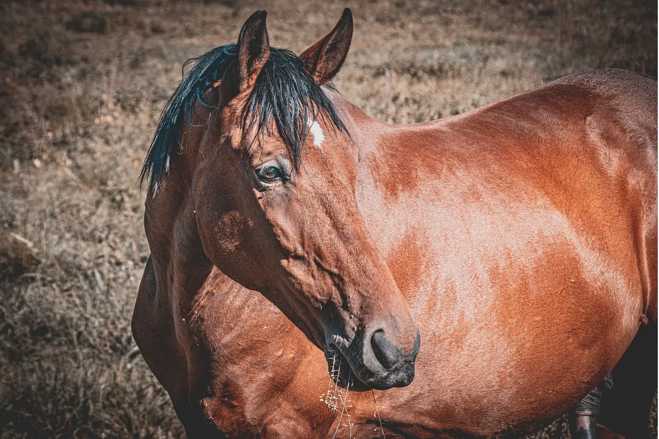
Horses have a 360 degree total visual field. This means that horses can see their tail with their heads pointed forward. Post-natally, a 65 degree frontal binocular view develops. Horses have a retina that is capable of detecting movement. The horse uses both eyes until the object is within 3-4 feet. If it cannot see with one eye, it will turn its head or lower itself to continue watching with the other eye. Horses have cones in their retina which indicate that they can see color in the form blues and reds.

OCULAR PROBLEMS IN FOAL
A newborn foal might have droopy eyes, low tear secretion and a round pupil. There may also be a reduced menace reflex, prominent lens sutures, and decreased corneal sensitivity. Entropion refers to an inward roll of the eyelid margin. The eyelid hairs rub against the cornea. This can occur in foals as a primary problem or secondary to dehydration and emaciation in “downer foals”. To prevent corneal ulceration in neonates, it can be fixed by placing sutures along the lid margin.
Common congenital eye defects are congenital cataracts in foals. Surgery is highly recommended. A common congenital defect is microphthalmos, or a small-sized eye. There may be a variety of lesions. Microphthalmic eye can be either visual or may be associated with other eye conditions that lead to blindness.
Uveitis and iridocyclitis in foals are usually secondary to severe illness. They may occur in one or both of the eyes. There may also be red cells, white cells, and proteins. In the southern United States, yearlings and foals of Thoroughbreds have been diagnosed with severe unilateral blinding fibrinous vision impairments secondary to plant toxins.
EYE DIAGNOSES AND THERAPY
Traumatic eyelid lesions
To prevent corneal dehydration and further injury, a rapid and accurate treatment of limb trauma is essential. The eyelids are very vascularized and have an incredible ability to heal and resist infection. They can also swell very rapidly. Due to their large blood supply, minimal debridement should be done. An eyelid “tag”, or pedicle flap, should not ever be removed as it can lead to corneal ulceration and exposure keratitis.
Horses are more likely to sustain upper eyelid damage than those with lower ones. This is because the horse’s upper lid moves more than the lower one. To preserve eyelid function, it is important to preserve the margin of the eyelid. Protect the repaired lesion from “self trauma” by using masks or hard cups.
Neoplasia in the lids
Eyelid melanomas can be found in grey horses. Percherons and Arabians are also at higher risk. Melanomas can be single or multi-faceted. Cimetidine, surgical excision, and/or cryotherapy are the options for treatment.
Sarcoids can be single or multiple tumors in the eyelids and periocular area of horses. The etiology of sarcoids could be due to retroviruses or papilloma virus. Flies could be capable of transferring sarcoid cells between horses to cause traumatic skin lesions in others. Horses can be aggressive in their sarcoid behavior depending on where they live. Mules seem to be affected by an aggressive form of the sarcoids. The immunotherapy for sarcoids can be done using reduced Mycobacterium bovis cell walls extracts, such as the immunostimulant Bacillus Calmette-Gaerin. Before using BCG, it is worth applying antipsoriasis creams and/or topical 5-fluorouracil (5FU) for two weeks. Sarcoids can also be treated with intralesional radiotherapy, cryotherapy, hyperthermia and carbon dioxide laser excision. There have been varying success rates when intralesional chemotherapeutics, such as 5-FU or Cisplatin, were used. Some sarcoids can be treated with homeopathic remedies and caustic chemical creams.
Squamous cell carcinoma is the most common eye and lid tumor in horses. This could be due to increased carcinogenesis, periocular pigmentation, and the ultraviolet (UV), component of solar radiation. SCC is most likely to be caused by the UV component. Horses are more likely to develop SCC if they are older than the mean age of diagnosis (11.1 +/- ). One report shows a 0.4-year increase in prevalence. Clydesdales, Belgians and other draft horses are most likely to have ocular SCC. Next, Appaloosas or Paints are more common than Appaloosas or Paints. Quarter Horses, Thoroughbreds, Arabians and Quarter Horses have the lowest prevalence. Ocular SCC is more common in palomino, white, and grey-white hair colors than in brown, black, and bay hair coats. After surgical excision of the equine eye ocular SCC, you should consider immunotherapy, cryotherapy, cryotherapy, radiofrequency hyperthermia and CO2 laser ablation, or intralesional chemotherapy.
DISEASES IN THE CORNEA
Equine corneal ulceration
Equine corneal uti is common in horses. It is a serious eye disease that requires prompt diagnosis, laboratory confirmation, and the appropriate medical and surgical treatment. There are many types of corneal ulcers. They can be simple, superficial, or deep-seated abrasions to the corneal epithelium. Horses with prominent eyes may be more susceptible to trauma corneal injuries. Horses may experience mild to moderate bacterial or fungal keratitis. However, serious complications should be treated promptly. No matter how minor or severe, corneal ulcers in horses must be treated immediately. Uveitis and corneal infection are serious concerns in any case of slight corneal ulceration. Uveitis and iridocyclitis can occur in any type of corneal ulcer. They must be treated to prevent vision loss.
Proteinases found in the tear film
Tear film proteinases are normally used to monitor and repair damaged cells and collagen due to regular wear and tear on the cornea. To prevent tissue from being too damaged, these enzymes are balanced with inhibitory substances. Excessive levels of these proteinsases may lead to rapid degeneration and destruction of collagen and other components in the stroma. This can cause keratomalacia (or corneal “melting”), which can be fatal.
Corneal sensitivity of foals and adult horses
The corneal sensation is essential for corneal healing. The cornea of an adult horse is more sensitive than other animals. Corneal touch threshold analysis showed that the corneas of hospitalized foals and adult horses were less sensitive than normal foals or adults. Sick neonates are more likely to develop corneal disease than healthy foals. This may partly explain why sick neonates with corneal disease are often without clinical signs.
Corneal healing for horses
The thickness of an equine cornea ranges from 1.0 to 1.5mm in the middle to 0.8mm at its periphery. The healing of large-diameter, noninfected corneal injuries is usually rapid and linear for five to 7 days. After that, it slows down. The increased activity of tear proteinase may cause slower healing in the second eye. Horses heal a corneal wound of 7 mm diameter that is not infected within 12 days (0.6 mm/day).
The horse corneal microenvironment
Horses live in a hostile environment. The cornea and conjunctiva are exposed to bacteria and fungal infections. The horse’s corneal epithelium is a strong barrier against the invasion and colonization of potentially pathogenic bacteria and fungi that are normally found on the cornea and conjunctiva. Defective corneal epithelium can allow bacteria and fungi to attach to the cornea, causing infection. Every corneal ulcer on a horse should be treated for infection. If a history of corneal injuries with vegetative material or prolonged corticosteroid and antibiotic therapy has not resulted in a significant improvement, it is important to suspect fungal involvement. Melting is a condition where excessive proteinase activity results in a liquefied, grayish -gelatinous appearance of the stroma at the margin of the ulcer. Horse corneas show a strong fibrovascular healing response. Horses have a unique corneal healing ability that is specific to their species. Horses suffering from painful eyes should have their corneas stained both with fluorescein dye or rose bengal dye. Fungal ulcers at the early stages of development will be negative for the fluorescein, but positive for rose bengal. First, corneal cultures should be taken. Then, corneal scrapings should be done for cytology. It is possible to have mixed bacterial and fungal infections.
Medical Therapy
To ensure complete treatment, it is important to carefully consider the therapy options once a corneal injury has been diagnosed. The first major thrust in controlling a corneal ulcer is medical therapy. However, it should be tempered with the careful use of additional surgical procedures. Modifying this intensive pharmacological treatment should be based on its effectiveness.
Antibiotics
Topically applied antibiotics, such as chloramphenicol, bacitracin-neomycin-polymyxin B, gentamicin, ciprofloxacin or tobramycin ophthalmic solutions may be utilized to treat bacterial ulcers. The frequency of medication can vary from q2h up to q8h. Cefazolin (55mg/ml), bacitracin, chloramphenicol and bacitracin are all effective against beta hemolytic Streptococcus. Topically, gentamicin-resistant Pseudomonas can be treated with ciprofloxacin (10 mg/ml), amikacin (10mg/ml), and polymyxin (0.25% IV solution).
Collagenolysis prevention
Extreme corneal inflammation due to bacterial infection (particularly, Pseudomonas, beta hemolytic Streptococcus), or fungal infection can cause rapid corneal liquefaction, and even perforation. Stromal collagenolysis and melting are caused by activation and/or production proteolytic enzymes. Serum is non-toxic, and it contains an alpha-2 macroglobulin that has antiproteinase activity. Topically administered serum can reduce the amount of tear film and corneal protease activity in horses with corneal ulcers. You can apply the serum topically as many times as you like, but it should be replaced every eight days.
Treat Uveitis
Atropine sulfate can be used to treat equine eye issues. Topically applied atropine (1%) can be used to stabilize the blood-aqueous membrane, reduce vascular protein leakage, minimize pain from ciliary muscles spasms, and reduce the likelihood of synechia formation through pupillary dilatation. Topically, Atropine can be used q4h to Q6h. The frequency of administration will decrease as soon as the pupil dilates. Topical atropine has been shown in horses to increase intestinal transit time, decrease and abolish intestinal sounds, as well as diminish normal myoelectric patterns within the small intestine. Some horses are more sensitive to atropine than others, and they may respond by showing signs of colic or prolonged intestinal transit time.
Flunixin meglumine (1 mg/kg IV, IM, or PO) are systemically administered NSAIDs. They can be taken orally or parenterally and have been shown to reduce uveal exudation in horses suffering from ulcers.
Uveitis can also be reduced by topical nonsteroidal anti-inflammatory drug (NSAIDs), such as profenol and flurbiprofen (BID to TID). Stall-return horses with corneal ulcers or secondary uveitis must be kept in a stable until they heal. Overexertion can lead to intraocular hemorhage or severe uveitis.
Conjunctival flaps
In equine ophthalmology, conjunctival flaps (or grafts) are often used to treat deep, melting, large, and perforated corneal injuries.
Unappropriate Therapy and Ulcers
Topical corticosteroids can encourage the growth of fungal and bacterial opportunists. They interfere with non-specific inflammation reactions and cell immunity. All forms of corticosteroid therapy are contraindicated for the treatment of corneal infection. If organisms are not removed from the corneal stroma, even topical corticosteroid injections can be fatal.
PLEASE READ THE FOLLOWING**
Corneal ulcers can be difficult to see even under proper lighting.
All red eyes or those with severe pain must be stained using fluorescein or rose bengal dyes.
* Slowly progressive and indolent courses often deceive the seriousness of an ulcer.
Corneal ulcers may quickly progress in horses to an eye tear.
Topical corticosteroids can cause corneal fluorescein staining.
* It is possible to have uveitis due to a corneal or stromal ulcer.
FUNGAL ULTRAVISORS IN HORSES
Fungi are common inhabitants of the horse environment and conjunctival microflora, but can be pathogenic after corneal injury. Fusarium and Aspergillus are all known causes of fungal keratomycosis in horses. Standardbreds seem to be more resistant to severe keratomycosis than Saddlebreds. The treatment can be quite long and may cause corneal scarring. These fungi are more susceptible to antifungal drugs than others in the following order: natamycin = miconazole, itraconazole, ketoconazole and fluconazole.
CORNEAL STROMAL ABSCESSES
Small epithelial ulcerative micropunctures can be used to inject microbes or debris into the corneal lumen. An epithelial cell adjacent to the micropuncture may divide and migrate to the small traumatic wound to encapsulate foreign bodies or infectious agents. This can lead to a corneal abscess. A fungal infection is more common than a bacterial one. The medical treatment consists of the aggressive use of systemic and topical antibiotics, topical atropine, and topical and systemsic NSAIDs. In abscesses close to Descemet’s membrane, deep lamellar or penetrating Keratoplasties are used. PK removes the sequestered microbial antibiotics, cyotokines, and toxins from the abscess.
CATARACTS IN HORSE
Cataracts, which are opacities in the lens, are the most common congenital ocular defect among foals. As horses age, they develop different degrees of blindness. Blindness is not usually associated with very small incipient lenses opacities. Blindness will increase as cataracts mature and become opaque.
Equine Cataract surgery
If the foal is in good health, there are no other ocular conditions and the foal can tolerate aggressive medical treatment.
The most effective technique for horses is phacoemulsification cataract surgery. After removing the anterior capsule, this extracapsular procedure uses a piezoelectric handpiece and an ultrasonic titanium needle. After the lens has emulsified, it is removed from the eye. Intraocular pressure is not affected. The posterior capsule, which is thin and fragile, is kept intact. Most horses recover quickly from phacoemulsification surgery. There is also less inflammation after the procedure. While the results of cataract surgery on foals by skilled veterinary ophthalmologists can be very good, they are not as good for adult horses suffering from cataracts due to ERU. Problem is that ERU can cause new blood vessels to form on the iris and anterior lenses capsule, which can lead to complications. Sometimes, severe hyphema and hemorhage cannot be stopped by the surgeon.
DISEASES OF UVEAL TRACT
Equine persistent uveitis (Periodic eye, moon blindness and iridocyclitis).
Equivine recurrent uveitis is a common cause for blindness in horses. It’s a group immune-mediated diseases with multiple causes. ERU is known for the recurrence and occurrence of anterior uveitis. In approximately 20% of cases, the disease is bilateral. Although the cause of ERU is clear to be immune-mediated, it remains unknown what the exact causes are. Hypersensitivity to infectious agents like Leptospirainterrogans is often suspected as a cause. In the U.S., you can request Leptospiral Titers for L. pomona and L. bratislava.
It is important to have positive titers of serovars that are greater than 1:400. The prognostic value of blindness in one or both eyes can be assessed using serology for Leptospira Pomona. Blindness in one or both eyes within 11 years after the first attack is common in seropositive Appaloosas (100%), seronegative Appaloosas (72%), seropositive Appaloosas (51%), and seropositive Non-Appaloosas (34%).
To determine if the uveitis may be caused by a corneal injury, a complete ophthalmic exam should be done. A corneal ulcer can prevent the use of topical corticosteroids. However, it is not recommended to use any nonsteroidal topical drugs. ERU can cause brain inflammation. ERU can lead to irreversible blindness. This is caused by retinal detachment or cataract formation.
ERU therapy
ERU treatment aims to prevent or minimize recurrences of uveitis attacks, preserve vision and decrease pain. It is difficult to determine the exact cause of each case and provide treatment. To maintain transparency of the ocular structures, treatment should be prompt and aggressive. Once clinical signs have subsided, medications should be gradually reduced in frequency. The treatment can last several weeks to months. Do not stop abruptly, as recurrences may occur.
Horses may need lifelong therapy.
ERU can be treated, although the outlook is not good. The Appaloosa dog seems to be most affected by the most severe cases.
To control intense intraocular inflammation, corticosteroids as well as nonsteroidal drugs are used. You can administer medication topically, either subconjunctivally or intramuscularly, and/or intravenously. Initial use should be made of dexamethasone or prednisolone. Subconjunctival corticosteroids can be used when frequent topical steroids are not feasible. Although systemic corticosteroids can be helpful in the treatment of severe cases of ERU that are refractory, they may cause laminitis. Nonsteroidal anti-inflammatory drug (NSAIDs) may have additive anti-inflammatory effects. They are effective in reducing intraocular inflammation if a corneal injury is present. ERU can be treated topically with Cyclosporine A. To control intraocular inflammation, flunixin meglumine and phenylbutazone are often used systemically. Some horses may not respond to these medications and need to be switched to another NSAID. Topical atropine reduces the risk of synechiae by inducing mydriasis. It also relieves some of ERU’s pain by relieving spasms of the ciliary muscles.
Surgical considerations in ERU
Vitrectomy is more effective in European warmbloods with ERU, than in Appaloosas in the U.S. It is not clear why. In both areas, cataracts are common. Postoperatively, retinal detachment may also be possible. Treatment of ERU may be possible with the use of intravitreal sustained release cyclosporine A implant.
RETINOPATHIES
Congenital stationary-night blindness
Congenital Stationary Nightblindness (CSNB), which is primarily found in the Appaloosa is inherited as a sex linked recessive trait. These cases are also reported in Standardbreds, Paso Finos, and Thoroughbreds. The clinical signs are visual impairment in dim lighting with normal vision in daylight, and behavioral uneasiness or unpredictability at night. CSNB is not a progressive condition, so its name. However, some cases of vision problems in the daytime can be noted. Normal ophthalmoscopic examinations are performed. Diagnosis can be made by clinical signs, breed, and electroretinogram (ERG). There is a decreased scotopic frequency and an increased scotopic b wave amplitude. Functional abnormalities in neurotransmission in the middle retina are believed to cause CSNB. This condition is not treatable, but animals with it should be avoided.
SUDDEN BLINDESS
Acute blindness can be caused by head or ocular trauma, ERU or glaucoma. Horses who are acutely blind can be extremely anxious, agitated and dangerous. Horses can adjust well to being blind, unilaterally or bilaterally, if they are allowed to. Many Internet sites offer information on caring for blind horses and other animals.
EYE DISEASES ASSOCIATED TO SPECIFIC HORSE BREEDS
APPALOOSA
1. Congenital stationary and night blindness (CSNB).
2. Congenital cataracts
3. Glaucoma
4. ERU
5. Optic disc colobomas
ARABIAN
1. Congenital cataracts
BELGIAN DRAFT HERRES
1. Secondary cataracts and aniridia
2. Cataracts
MORGAN
1. Cataracts: Nuclear, bilateral, symmetrical and non-progressive
QUARTER HERSE
1. Congenital cataracts
2. Entropion
ROCKY MOUNTAIN HOUSE (chocolate coat most commonly affected). Anterior segment dysgenesis is collectively known as the cornea, iris, and ciliary body lesion.
1. Congenital miosis and corpora-nigra and Irhyplasia
2. Macrocornea
3. Ciliary cysts
4. Cataract, Lens Luxation
5. Retinal Detachment, Retinal Dysplasia
THOROUGHBRED
1. Congenital cataracts
2. Multiple ocular defects can lead to microphthalmia.
3. In some cases, retinal detachments can cause retinal dysplasia.
4. Entropion
5. Progressive retinal atrophy
COLOR DILUTED BREEDS
1. Photophobia – Iridal hypoplasia
STANDARDBREDS
1. Retinal detachments
2. Congenital Stationary Nightblindness (CSNB).
PASOFINO
1. CSNB
2. Glaucoma
AMERICAN ADDLEBRED
1. Cataracts
2. Keratomycosis aggressive
WARM BLOOD
1. Glaucoma
2. ERU
MINIATURE HORSES
1. Cataracts
MULES
1. Sarcoids who are aggressive